Fish Heart Chambers Diagram - Anatomy Of Larval Fish A Representative General Larval Forms Download Scientific Diagram : The heart is not a neural because endocardial connective tissue in the portal heart of eptatretus burgeri contains nerve bundles.
Fish Heart Chambers Diagram - Anatomy Of Larval Fish A Representative General Larval Forms Download Scientific Diagram : The heart is not a neural because endocardial connective tissue in the portal heart of eptatretus burgeri contains nerve bundles.. Getting a 3 chambered heart from a 2 chambered heart:t: 3 and 4 chambered hearts are similar because they both have an interior circuit or double circulation. 2 chambers, amphibians and reptiles: The ventricle, however, remains as a single chamber. In fish, the system has only one circuit, with the blood being pumped through the capillaries of the gills and on to the capillaries of the body tissues.
Of the vertebrates, or animals with a backbone, fish have the simplest type of heart and is considered the next step in the evolutionary chain. Look at the diagrams and follow in your shark the passage of water in the mouth and spiracles (which have a one way valve) and through the five gill slits. A schematic diagram of the three main phases of filling and emptying of four cardiac chambers of the fish heart. An atrium and a ventricle. Human heart • four chambers • double circuit • oxygen poor blood is pumped from the right side of the heart to the lungs where it is oxygenated.
Circulatory Systems from www1.biologie.uni-hamburg.de Blood then is returned to the heart. The basic vertebrate heart, such as occurs in fish, has two. While it is a closed circulatory system, it has only two chambers. Given a 2 chambered heart, experts do not know when, how, or in what lineage the alleged transition from the 2 chamber fish heart to the 3 chambered Human heart • four chambers • double circuit • oxygen poor blood is pumped from the right side of the heart to the lungs where it is oxygenated. The wall of these chambers consists of the same components as in mammals — the endocardium as the inside lining, the middle myocardium and the outer epicardium. In comparison, the human heart has two separate ventricles and two separate atria. A fish's heart has four chambers.
Their heart consists of one auricle or atrium, and one ventricle.
They only have single circulation of blood and definitely possess the lowest pressure and least advanced heart. Egg are then released into the oviduct. The atrium receives blood from the veins, and the ventricle pumps blood to the gills for gas exchange, similar to the ventricle in frogs. There heart circulates blood through the body at a. The heart is not a neural because endocardial connective tissue in the portal heart of eptatretus burgeri contains nerve bundles. This diagram shows the female reproductive system. What conclusions can one draw from the above diagram? The venous side of the heart is preceded by an enlarged chamber called the sinus venosus. A fish's heart has four chambers. The deoxygenated blood enters through the sinus venosus and into the atrium. To study these features examine the models of hearts and aortic arches displayed in the lab, the illustrations in your lab and textbook and what you have learned about the dogfish, mudpuppy, cat and rat. Oxygen rich blood then flows back to the left side of the heart and is pumped from there through the rest of the body. This is known as single cycle circulation.
Human heart • four chambers • double circuit • oxygen poor blood is pumped from the right side of the heart to the lungs where it is oxygenated. The fish heart has one ventricle and one atrium. The heart pumps the blood in a single loop throughout the body. This single circuit is known as systemic circulation. Diagram's of the circulatory system.
Fish Heart Chamber File Fish Heart Schematic Png Wikimedia Commons from lh4.googleusercontent.com When the pharynx is filled, the mouth closes and the gill chambers expand and fill with water. This is known as single cycle circulation. There heart circulates blood through the body at a. An auricle is the chamber of the heart where blood is received from the body. In fish, the system has only one circuit, with the blood being pumped through the capillaries of the gills and on to the capillaries of the body tissues. The deoxygenated blood enters through the sinus venosus and into the atrium. The right atrium receives blood from the veins and pumps it to the right ventricle. The right atrium tends to be larger than the left in most birds.
Their heart consists of one auricle or atrium, and one ventricle.
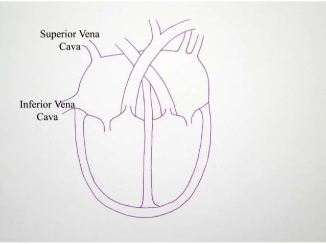
The arterial side of the heart is followed by a thickened muscular cavity called the bulbus arteriosus. The heart of fish is therefore only a single pump (consisting of two chambers). The top is called the atrium and the bottom chamber is called the ventricle. The sinus venosus (before the ventricle) and the bulbus arteriosus (after the atrium). Their heart consists of four parts: The atrium and ventricle constitute the classic '2 chambered fish heart'. There is thus only one atrium and one ventricle in the mature fish heart. (a) (i) compare the heart rate of the fit person with the heart rate of the unfit person from 5 to 15 minutes. Diagram's of the circulatory system. The fish heart has one ventricle and one atrium. But unlike us, the chambers of their heart are not all muscular and are not so built into a single organ. Oxygen rich blood then flows back to the left side of the heart and is pumped from there through the rest of the body. Human, fish and reptile heart.
(b) the atrium contracts (as shown by the small arrows), further expanding and completing the filling of the ventricle. When the pharynx is filled, the mouth closes and the gill chambers expand and fill with water. The heart, as well as the entire circulatory system, are unevolvable in a step by step manner. The two lateral oviducts, one for each ovary, join at the common oviduct. (a) (i) compare the heart rate of the fit person with the heart rate of the unfit person from 5 to 15 minutes.
Human Fish And Reptile Heart from image.slidesharecdn.com They only have single circulation of blood and definitely possess the lowest pressure and least advanced heart. 2 chambers, amphibians and reptiles: The wall of these chambers consists of the same components as in mammals — the endocardium as the inside lining, the middle myocardium and the outer epicardium. The venous side of the heart is preceded by an enlarged chamber called the sinus venosus. But now in addition, the single atrium becomes divided into two. The blood moves from the gills throughout the rest of the fish's body. A schematic diagram of the three main phases of filling and emptying of four cardiac chambers of the fish heart. The deoxygenated blood enters through the sinus venosus and into the atrium.
The four chambers of the heart are completely divided into two atria and two ventricles.
Oxygen rich blood then flows back to the left side of the heart and is pumped from there through the rest of the body. The ventricle, however, remains as a single chamber. A bony fish's heart has two chambers: A bony fishes heart has two chambers: The heart pumps the blood in a single loop throughout the body. What conclusions can one draw from the above diagram? The sinus venosus (before the ventricle) and the bulbus arteriosus (after the atrium). They only have single circulation of blood and definitely possess the lowest pressure and least advanced heart. A schematic diagram of the three main phases of filling and emptying of four cardiac chambers of the fish heart. Diagram's of the circulatory system. But unlike us, the chambers of their heart are not all muscular and are not so built into a single organ. The right atrium tends to be larger than the left in most birds. In comparison, the human heart has two separate ventricles and two separate atria.


Posting Komentar
0 Komentar